랑게르한스섬
랑게르한스섬(islets of Langerhans, pancreatic islets), 또는 이자 섬은 이자에 있는 내분비 조직이다. 1869년 해부병리학자 파울 랑게르한스에 의해 발견되었다.[1] 이자 섬은 이자 부피의 1~2%를 차지하며 이자에 들어오는 10~15%의 혈류가 통과한다.[2][3] 이자 섬은 이자 전체에 걸쳐 조밀하게 분포되어 있으며, 포도당의 대사에 중요한 역할을 한다.[4]
표피의 랑게르한스 세포와는 다른 조직이다.
구조 편집
건강한 어른의 경우 약 3백만 개의 이자 섬이 이자 전체에 걸쳐 조밀한 섬 형태로 분포하고 있으며, 각각 0.1 mm (109 µm)의 지름을 가진다.[5]:914 각각의 섬은 이자의 다른 부분에 걸쳐 엮여 있는 섬유상 연결 조직과 연속적으로 연결된 섬유성 연결 조직 캡슐로 이자의 다른 부분과 구분된다.[5]:914
미세해부학 편집
이자 섬에서 호르몬을 생성되어 바로 혈류로 분비하는 세포는 최소 5가지가 알려져 있다. 쥐의 이자 섬에서 세포의 구분은 다음과 같이 구별된다:[6]
- 알파세포: 글루카곤 생성 (전체 이자섬 세포의 20%)
- 베타세포: 인슐린과 아밀린 생성 (≈70%)
- 델타세포: 소마토스타틴 생성 (<10%)
- 입실론세포: 그렐린 생성 (<1%)
- PP 세포 (감마세포 또는 F세포): 이자 단백질 생성 (<5%)
이자 섬의 세포 구조는 종마다 다르다는 것이 알려져있다.[7][8][9] 예시로 설치류의 이자 섬은 인슐린을 생성하는 베타 세포가 중심부에 주로 분포하며 알파, 델타, PP 세포가 주변부에 적게 분포하나, 인간의 이자 섬은 알파 세포와 베타 세포가 전체 이자 섬에 비슷한 비율로 존재한다.[7][9]
이자 섬은 자가분비와 측분비 신호전달을 통해 서로에게 영향을 줄 수 있으며, 베타 세포는 여섯 개에서 일곱 개 정도의 다른 베타 세포와 전기적으로 연결되어 있다.[10]
-
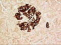
 염색된 이자 섬
 -
 이자 섬, 알파 세포
 -
 이자 섬, 베타 세포


기능 편집
이자 섬의 측분비 되먹임 계는 다음과 같은 구조를 가진다:[11]
- 포도당/인슐린: 베타 세포의 활성화, 알파 세포의 억제.
- 글리코겐/글루카곤: 알파 세포의 활성화를 통해 베타 세포와 델타 세포를 활성화.
- 소마토스타틴: 알파 세포와 베타 세포를 억제.
많은 수의 G 단백질 연결 수용체가 이자 섬에서의 인슐린, 글루카곤 그리고 소마토스타틴의 분비를 조절한다.[12] 일부 G 단백질 연결 수용체는 2형 당뇨 치료제의 타겟으로 작용한다 (GLP-1 수용체 길항제, DPPIV 억제제).
-
 췌장 단백질 항체로 염색한 쥐의 이자 섬
 -
 인슐린 항체로 염색한 쥐의 이자 섬
 -
 글루카곤 항체로 염색된 쥐의 이자 섬


전기적 활성 편집
이자 섬의 전기 활성은 패치 클램프 기술을 이용해 연구 되어 왔다. 이를 통해 건강한 이자 섬에서의 세포 거동이 그렇지 못한 이자 섬에서의 세포 거동과 다르다는 것이 밝혀졌다.[13]
임상적 의미 편집
당뇨 편집
이자 섬의 베타 세포는 인슐린을 분비하며, 당뇨에서 중요한 역할을 한다. 당뇨병 환자에서 베타 세포는 자가 면역 작용에 의해 파괴된다고 알려져 있다. 반대로 베타 세포가 파괴되지 않고 단지 기능을 하지 않는다는 보고도 있다.
장기이식 편집
제 1형 당뇨병 환자의 이자 섬 베타 세포는 자가 면역 반응에 의해 선택적으로 파괴된다. 이에 베타 세포의 기능을 회복하기 위해 이자 이식이나 인공 이자를 대신할 이자 섬 이식이 활발히 연구되고 있다.[14][15] 이자 섬 이식은 1970년대 당뇨병 치료를 위한 치료법으로 떠올랐으며 지난 30년간 꾸준히 발전했다.[16] 최근의 임상 시험에 따르면 장기기증자의 이자 섬을 제 1형 당뇨병 환자에게 이식 한 후 인슐린 독립성과 개선된 대사 조절을 재현성있게 얻을 수있는 것으로 나타났다.[15]
제 1형 당뇨병에 대한 이자 섬 이식은 현대 수여자의 면역 거부를 예방하기 위한 면역 억제가 필요하다.[17]
성체 줄기 세포 또는 전구 세포로부터 유래된 인슐린 생성 세포가 장기 기증의 부족을 극복하기 위한 대안책으로 떠오르고 있다. 재생 의학이 빠르게 발전함에 따라 가까운 미래에 해당 기술이 개발될 것으로 보인다. 한편, 제 1형 당뇨가 이자의 베타 세포에 대한 자가 면역에 의한 파괴 때문이므로, 적절하고 안전한 면역 중재와 베타 세포 재생 접근법을 결합한 순차적 접근법이 필요하다.[18] 알파 세포가 베타 세포로 분화 할 수 있다는 것도 입증되었다. 건강한 사람과 당뇨병 환자, 쥐의 알파 세포 모두 자발적으로 베타 세포로 전환될 수 있어 미래의 베타 세포 치료법으로 예상된다.[19] 실제로, 섬 형태와 내분비 분화가 직접적으로 관련되어 있음이 밝혀졌다.[20] 내분비 선조 세포는 응집력에 의해 새싹 같은 섬 전구체 또는 "반도"를 형성함으로써 분화되는데, 여기서 알파 세포는 반도 외층을 구성하고 베타 세포는 그 아래에 형성된다.
이미지 편집
-
 이자 섬
이자 섬 -
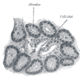 개 이자 일러스트. 250x.
 -
 쥐 이자(위)와 사람 이자(아래)의 구조적 차이. 왼쪽은 이자의 복측, 오른쪽은 이자의 배측. 다른 세포는 색으로 구분된다. 설치류의 이자 섬은 사람의 것과 다른 인슐린 코어를 보인다.


관련문서 편집
출처 편집
- ↑ Langerhans P (1869). “Beitrage zur mikroscopischen anatomie der bauchspeichel druse”. 《Inaugural-dissertation. Berlin: Gustav Lange》.
- ↑ Barrett KE, Boitano S, Barman SM, Brooks HL (2009년 7월 22일). 《Ganong's review of medical physiology》 23판. McGraw Hill Medical. 316쪽. ISBN 978-0-07-160568-7.
- ↑ Functional Anatomy of the Endocrine Pancreas
- ↑ Pour, Parviz M.; Standop, Jens; Batra, Surinder K. (January 2002). “Are islet cells the gatekeepers of the pancreas?”. 《Pancreatology》 2 (5): 440–448. doi:10.1159/000064718. PMID 12378111.
- ↑ 가 나 Sleisenger, edited by Mark Feldman, Lawrence S. Friedman, Lawrence J. Brandt; consulting editor, Marvin H. (2009). 《Sleisenger & Fordtran's gastrointestinal and liver disease pathophysiology, diagnosis, management》 9판. St. Louis, Missouri: MD Consult. ISBN 978-1-4160-6189-2.
- ↑ Elayat AA; el-Naggar MM; Tahir M; Bassam dahrouj (1995). “An immunocytochemical and morphometric study of the rat pancreatic islets”. 《Journal of Anatomy》. 186. (Pt 3) (Pt 3): 629–37. PMC 1167020. PMID 7559135.
- ↑ 가 나 Brissova M, Fowler MJ, Nicholson WE, Chu A, Hirshberg B, Harlan DM, Powers AC (2005). “Assessment of human pancreatic islet architecture and composition by laser scanning confocal microscopy”. 《Journal of Histochemistry and Cytochemistry》 53 (9): 1087–97. doi:10.1369/jhc.5C6684.2005. PMID 15923354.
- ↑ Ichii H, Inverardi L, Pileggi A, Molano RD, Cabrera O, Caicedo A, Messinger S, Kuroda Y, Berggren PO, Ricordi C (2005). “A novel method for the assessment of cellular composition and beta-cell viability in human islet preparations”. 《American Journal of Transplantation》 5 (7): 1635–45. CiteSeerX 10.1.1.578.5893. doi:10.1111/j.1600-6143.2005.00913.x. PMID 15943621.
- ↑ 가 나 Cabrera O, Berman DM, Kenyon NS, Ricordi C, Berggren PO, Caicedo A (2006). “The unique cytoarchitecture of human pancreatic islets has implications for islet cell function”. 《Proceedings of the National Academy of Sciences of the United States of America》 103 (7): 2334–9. Bibcode:2006PNAS..103.2334C. doi:10.1073/pnas.0510790103. ISSN 1091-6490. PMC 1413730. PMID 16461897.
- ↑ Kelly, Catriona; McClenaghan, Neville H.; Flatt, Peter R. (2011). “Role of islet structure and cellular interactions in the control of insulin secretion”. 《Islets》 3 (2): 41–47. doi:10.4161/isl.3.2.14805. PMID 21372635.
- ↑ Wang, Michael B.; Bullock, John; Boyle, Joseph R. (2001). 《Physiology》. Hagerstown, MD: Lippincott Williams & Wilkins. 391쪽. ISBN 978-0-683-30603-3.
- ↑ "An atlas and functional analysis of G-protein coupled receptors in human islets of Langerhans.Amisten S, Salehi A, Rorsman P, Jones PM, Persaud SJ., Pharmacol Ther. 2013 May 18. PMID 23694765
- ↑ Pérez-Armendariz M, Roy C, Spray DC, Bennett MV (1991). “Biophysical properties of gap junctions between freshly dispersed pairs of mouse pancreatic beta cells”. 《Biophysical Journal》 59 (1): 76–92. Bibcode:1991BpJ....59...76P. doi:10.1016/S0006-3495(91)82200-7. PMC 1281120. PMID 2015391.
- ↑ Meloche RM (2007). “Transplantation for the treatment of type 1 diabetes”. 《World Journal of Gastroenterology》 13 (47): 6347–55. doi:10.3748/wjg.13.6347. PMC 4205453. PMID 18081223.
- ↑ 가 나 Hogan A, Pileggi A, Ricordi C (2008). “Transplantation: current developments and future directions; the future of clinical islet transplantation as a cure for diabetes”. 《Frontiers in Bioscience》 13 (13): 1192–205. doi:10.2741/2755. PMID 17981623.
- ↑ Piemonti L, Pileggi A (2013). “25 Years of the Ricordi Automated Method for Islet Isolation”. 《CellR4》 1 (1): 8–22.
- ↑ Chatenoud L (2008). “Chemical immunosuppression in islet transplantation—friend or foe?”. 《New England Journal of Medicine》 358 (11): 1192–3. doi:10.1056/NEJMcibr0708067. ISSN 0028-4793. PMID 18337609.
- ↑ Pileggi A, Cobianchi L, Inverardi L, Ricordi C (2006). “Overcoming the challenges now limiting islet transplantation: a sequential, integrated approach”. 《Annals of the New York Academy of Sciences》 1079 (1): 383–98. Bibcode:2006NYASA1079..383P. doi:10.1196/annals.1375.059. ISSN 0077-8923. PMID 17130583.
- ↑ van der Meulen, T.; Mawla, A.M.; DiGruccio, M.R.; Adams, M.W.; Nies, V.; Dolleman, S.; Liu, S.; Ackermann, A.M.; Caceres, E.; Hunter, A.E.; Kaestner, K.H.; Donaldson, C.J.; Huising, M.O. (2017). “Virgin Beta Cells Persist throughout Life at a Neogenic Niche within Pancreatic Islets”. 《Cell Metabolism》 25 (4): 911–926. doi:10.1016/j.cmet.2017.03.017. PMID 28380380.
- ↑ Sharon, N.; Chawla, R.; Mueller, J.; Vanderhooft, J.; Whitehorn, L.J.; Rosenthal, B.; Gürtler, M.; Estanboulieh, R.R.; Shvartsman, D.; Gifford, D.K.; Trapnell, C.; Melton, D. (2019). “A Peninsular Structure Coordinates Asynchronous Differentiation with Morphogenesis to Generate Pancreatic Islets”. 《Cell》 176 (4): 790–804.e13. doi:10.1016/j.cell.2018.12.003. ISSN 0092-8674. PMC 6705176. PMID 30661759.
외부 링크 편집
- Pancreas at the Human Protein Atlas