파일:Mitochondria, mammalian lung - TEM.jpg
최대 해상도입니다.
Mitochondria,_mammalian_lung_-_TEM.jpg (640 × 480 픽셀, 파일 크기: 96 KB, MIME 종류: image/jpeg)
파일 설명
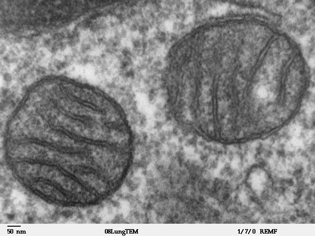
| 설명 |
Transmission electron microscope image of a thin section cut through an area of mammalian lung tissue. The high magnification image shows two mitochondria. JEOL 100CX TEM |
| 출처 | |
| 저자 | Louisa Howard |
| 저작권 (이 파일을 인용하기) |
PD |
라이선스
| 이 작품은 저작자인 Louisa Howard에 의해 퍼블릭 도메인으로 공개된 작품입니다. 이 공개 선언은 전 세계적으로 유효합니다. 만약 저작권의 포기가 법률적으로 가능하지 않은 경우, Louisa Howard은 이 작품을 법적으로 허용되는 한도 내에서 누구나 자유롭게 어떤 목적으로도 제한 없이 사용할 수 있도록 허용합니다.
|
파일 역사
날짜/시간 링크를 클릭하면 해당 시간의 파일을 볼 수 있습니다.
| 날짜/시간 | 섬네일 | 크기 | 사용자 | 설명 | |
|---|---|---|---|---|---|
| 현재 | 2008년 5월 17일 (토) 00:09 | | 640 × 480 (96 KB) | Vojtěch Dostál | Reverted to version as of 15:37, 5 October 2006, my fault |
| 2008년 5월 16일 (금) 21:51 |  | 640 × 433 (84 KB) | Vojtěch Dostál | {{Information |Description=Transmission electron microscope image of a thin section cut through an area of mammalian lung tissue. The high magnification image shows a mitochondria. JEOL 100CX TEM |Source= * http://remf.dartmouth.edu/imagesindex.html * h | |
| 2008년 5월 16일 (금) 21:47 |  | 640 × 453 (86 KB) | Vojtěch Dostál | {{Information |Description=Transmission electron microscope image of a thin section cut through an area of mammalian lung tissue. The high magnification image shows a mitochondria. JEOL 100CX TEM |Source= * http://remf.dartmouth.edu/imagesindex.html * h | |
| 2006년 10월 6일 (금) 00:37 |  | 640 × 480 (96 KB) | Patho | {{Information |Description=Transmission electron microscope image of a thin section cut through an area of mammalian lung tissue. The high magnification image shows a mitochondria. JEOL 100CX TEM |Source= * http://remf.dartmouth.edu/imagesindex.html * h |
이 파일을 사용하는 문서
다음 문서 1개가 이 파일을 사용하고 있습니다:
이 파일을 사용하고 있는 모든 위키의 문서 목록
다음 위키에서 이 파일을 사용하고 있습니다:
- ar.wikipedia.org에서 이 파일을 사용하고 있는 문서 목록
- az.wiktionary.org에서 이 파일을 사용하고 있는 문서 목록
- be.wikipedia.org에서 이 파일을 사용하고 있는 문서 목록
- bg.wikipedia.org에서 이 파일을 사용하고 있는 문서 목록
- bn.wikipedia.org에서 이 파일을 사용하고 있는 문서 목록
- br.wikipedia.org에서 이 파일을 사용하고 있는 문서 목록
- bs.wikipedia.org에서 이 파일을 사용하고 있는 문서 목록
- ca.wikipedia.org에서 이 파일을 사용하고 있는 문서 목록
- cdo.wikipedia.org에서 이 파일을 사용하고 있는 문서 목록
- da.wikipedia.org에서 이 파일을 사용하고 있는 문서 목록
- de.wikibooks.org에서 이 파일을 사용하고 있는 문서 목록
- el.wikipedia.org에서 이 파일을 사용하고 있는 문서 목록
- en.wikipedia.org에서 이 파일을 사용하고 있는 문서 목록
- en.wikibooks.org에서 이 파일을 사용하고 있는 문서 목록
- en.wikiversity.org에서 이 파일을 사용하고 있는 문서 목록
- User:Jtwsaddress42/Projects/Project 1
- User:Jtwsaddress42/Projects/Project 1/Parts
- User:Jtwsaddress42/Projects/Project 1/Parts/Part 3
- User:Jtwsaddress42/Projects/Project 1/Chapters/Chapter 10
- User:Jtwsaddress42/Projects/Project 1/Sections/Chapter 10/Phase II - The Oxygen Crisis and the Rise of the Aerobic Bioshphere (1.9-0.95 bya)
- User:Jtwsaddress42/Clade
- User:Jtwsaddress42/Clade/Gracilicutes to Proteobacteria
- en.wiktionary.org에서 이 파일을 사용하고 있는 문서 목록
- es.wikipedia.org에서 이 파일을 사용하고 있는 문서 목록
- et.wikipedia.org에서 이 파일을 사용하고 있는 문서 목록
- eu.wikipedia.org에서 이 파일을 사용하고 있는 문서 목록
- ext.wikipedia.org에서 이 파일을 사용하고 있는 문서 목록
- fa.wikipedia.org에서 이 파일을 사용하고 있는 문서 목록
- fr.wikipedia.org에서 이 파일을 사용하고 있는 문서 목록
이 파일의 더 많은 사용 내역을 봅니다.