파일:Mycobacterium tuberculosis 8438 lores.jpg
Mycobacterium_tuberculosis_8438_lores.jpg (700 × 475 픽셀, 파일 크기: 49 KB, MIME 종류: image/jpeg)
파일 설명
| 설명 |
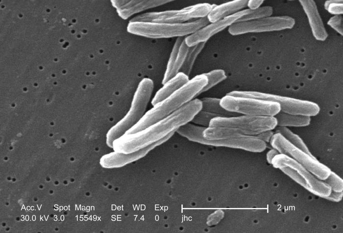
English: Under a high magnification of 15549x, this scanning electron micrograph (SEM) depicted some of the ultrastructural details seen in the cell-wall configuration of a number of Gram-positive Mycobacterium tuberculosis bacteria. As an obligate aerobic organism, M. tuberculosis can only survive in an environment containing oxygen. This bacterium ranges in length between 2-4 microns, with a width of 0.2-0.5 microns. See PHIL 9997 for a colorized version of this image.
TB bacteria become active, and begin to multiply, if the immune system can't stop them from growing. The bacteria attack the body and destroy tissue. If in the lungs, the bacteria can actually create a hole in the lung tissue. Some people develop active TB disease soon after becoming infected, before their immune system can fight off the bacteria. Other people may get sick later, when their immune system becomes weak for another reason. Babies and young children often have weak immune systems. People infected with HIV, the virus that causes AIDS, have very weak immune systems. Other people can have weak immune systems, too, especially people with any of these conditions: substance abuse; diabetes mellitus; silicosis; cancer of the head or neck; leukemia or Hodgkin's disease; severe kidney disease; low body weight; certain medical treatments (such as corticosteroid treatment or organ transplants); specialized treatment for rheumatoid arthritis, or Crohn's disease.Français : Mycobacterium tuberculosis grossi 15 549 fois.
Español: Mycobacterium tuberculosis ampliado a 15549x.
中文:掃描電子顯微鏡下的結核桿菌.
Suomi: Mycobacterium tuberculosis 15549-kertaisena suurennoksena.
Čeština: Bakterie Mycobacterium tuberculosis, původce TBC.
Magyar: Mycobacterium tuberculosis.
한국어: 결핵균의 전자현미경 사진.
Kurdî: Girtineke elektronmîkroskobîk a bakteriyên tûberkûlozê pêk tînin.
Afrikaans: 'n Skanderende mikrograaf van Mycobacterium tuberculosis.
粵語: 掃描電子顯微鏡下嘅結核桿菌. |
||
| 날짜 | |||
| 출처 |
|
||
| 저자 |
|
||
| 저작권 (이 파일을 인용하기) |
PD-USGov-HHS-CDC English: This image is in the public domain and thus free of any copyright restrictions. As a matter of courtesy, we request that the content provider be credited and notified in any public or private usage of this image. |
||
| 다른 버전 |
이 파일은 다음으로 파생됨: IRG activation following pathogen entry .jpg
|

라이선스
This image is a work of the Centers for Disease Control and Prevention, part of the United States Department of Health and Human Services, taken or made as part of an employee's official duties. As a work of the U.S. federal government, the image is in the public domain.
|
파일 역사
날짜/시간 링크를 클릭하면 해당 시간의 파일을 볼 수 있습니다.
| 날짜/시간 | 섬네일 | 크기 | 사용자 | 설명 | |
|---|---|---|---|---|---|
| 현재 | 2006년 4월 19일 (수) 04:45 | | 700 × 475 (49 KB) | Patho | {{Information| |Description= ID#: 8438 Description: Under a high magnification of 15549x, this scanning electron micrograph (SEM) depicted some of the ultrastructural details seen in the cell wall configuration of a number of Gram-positive Mycobacterium t |
이 파일을 사용하는 문서
다음 문서 1개가 이 파일을 사용하고 있습니다:
이 파일을 사용하고 있는 모든 위키의 문서 목록
다음 위키에서 이 파일을 사용하고 있습니다:
- af.wikipedia.org에서 이 파일을 사용하고 있는 문서 목록
- ar.wikipedia.org에서 이 파일을 사용하고 있는 문서 목록
- ast.wikipedia.org에서 이 파일을 사용하고 있는 문서 목록
- ca.wikipedia.org에서 이 파일을 사용하고 있는 문서 목록
- cs.wikipedia.org에서 이 파일을 사용하고 있는 문서 목록
- de.wikipedia.org에서 이 파일을 사용하고 있는 문서 목록
- de.wikibooks.org에서 이 파일을 사용하고 있는 문서 목록
- de.wikinews.org에서 이 파일을 사용하고 있는 문서 목록
- en.wikinews.org에서 이 파일을 사용하고 있는 문서 목록
- es.wikipedia.org에서 이 파일을 사용하고 있는 문서 목록
- eu.wikipedia.org에서 이 파일을 사용하고 있는 문서 목록
- ext.wikipedia.org에서 이 파일을 사용하고 있는 문서 목록
- fi.wikipedia.org에서 이 파일을 사용하고 있는 문서 목록
- fr.wikipedia.org에서 이 파일을 사용하고 있는 문서 목록
- fr.wiktionary.org에서 이 파일을 사용하고 있는 문서 목록
- fy.wikipedia.org에서 이 파일을 사용하고 있는 문서 목록
- gd.wikipedia.org에서 이 파일을 사용하고 있는 문서 목록
- hi.wikipedia.org에서 이 파일을 사용하고 있는 문서 목록
- hu.wikipedia.org에서 이 파일을 사용하고 있는 문서 목록
- kk.wikipedia.org에서 이 파일을 사용하고 있는 문서 목록
- ku.wikipedia.org에서 이 파일을 사용하고 있는 문서 목록
- lt.wikipedia.org에서 이 파일을 사용하고 있는 문서 목록
- lv.wikipedia.org에서 이 파일을 사용하고 있는 문서 목록
- no.wikipedia.org에서 이 파일을 사용하고 있는 문서 목록
- oc.wikipedia.org에서 이 파일을 사용하고 있는 문서 목록
- pl.wikipedia.org에서 이 파일을 사용하고 있는 문서 목록
- ro.wikipedia.org에서 이 파일을 사용하고 있는 문서 목록
- ru.wikipedia.org에서 이 파일을 사용하고 있는 문서 목록
- scn.wikipedia.org에서 이 파일을 사용하고 있는 문서 목록
- tr.wikipedia.org에서 이 파일을 사용하고 있는 문서 목록
이 파일의 더 많은 사용 내역을 봅니다.