파일:Yellow mite (Tydeidae), Lorryia formosa.jpg
미리 보기 크기: 800 × 545 픽셀 다른 해상도: 320 × 218 픽셀 | 640 × 436 픽셀 | 1,024 × 697 픽셀 | 1,280 × 871 픽셀 | 2,350 × 1,600 픽셀
원본 파일 (2,350 × 1,600 픽셀, 파일 크기: 2.22 MB, MIME 종류: image/jpeg)
파일 설명
| 설명 |
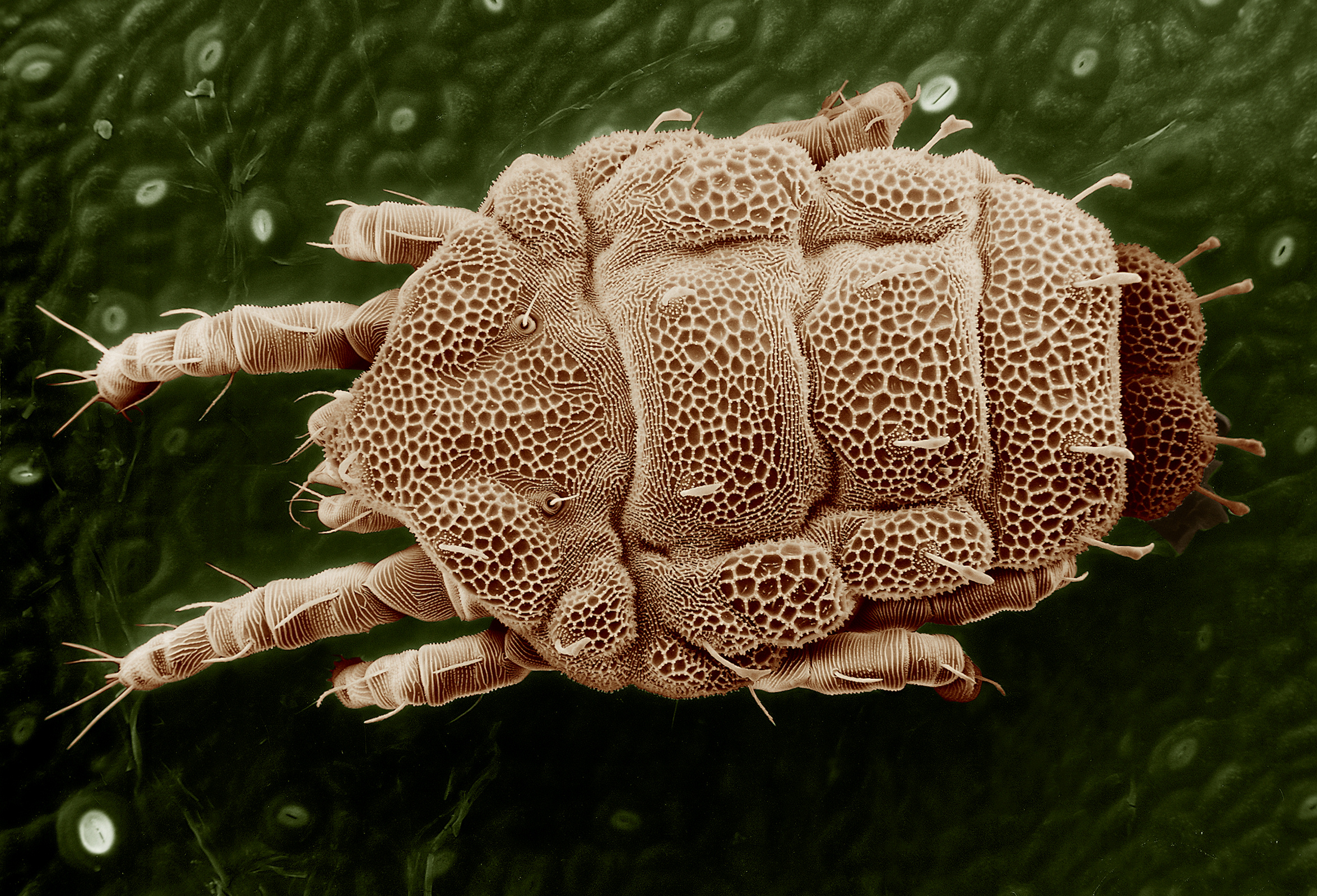
English: This is an overhead view of the Lorryia formosa mite. Magnified about 200×. Scanning electron microscopy allows mites to be viewed from different angles. Photo by Eric Erbe; digital colorization by Chris Pooley.
فارسی: نگارهای ثبتشده از بالاسر از هیره زرد. این نگاره بهوسيلهٔ لنزی ۲۰۰ برابری ثبت شدهاست. |
||
| 날짜 | 2008년 1월 28일 (Exif 데이터에서 가져옴) | ||
| 출처 |
|
||
| 저자 | Photo by Eric Erbe; digital colorization by Chris Pooley |
|

,_Lorryia_formosa.jpg?uselang=ko)
,_Lorryia_formosa.jpg)
,_Lorryia_formosa.jpg)
라이선스
| This image is in the public domain because it contains materials that originally came from the Agricultural Research Service, the research agency of the United States Department of Agriculture.
|
파일 역사
날짜/시간 링크를 클릭하면 해당 시간의 파일을 볼 수 있습니다.
| 날짜/시간 | 섬네일 | 크기 | 사용자 | 설명 | |
|---|---|---|---|---|---|
| 현재 | 2008년 1월 29일 (화) 07:39 | | 2,350 × 1,600 (2.22 MB) | Lycaon | noise processed |
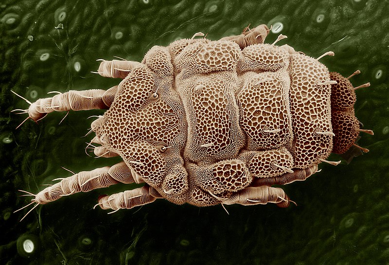
| 2005년 5월 5일 (목) 08:16 |  | 2,400 × 1,634 (3.86 MB) | Brian0918 | This is an overhead view of the Lorryia formosa mite. Magnified about 200x. Scanning electron microscopy allows mites to be viewed from different angles. Photo by Eric Erbe; digital colorization by Chris Pooley. http://www.ars.usda.gov/is/AR/archive/oct0 |
이 파일을 사용하는 문서
다음 문서 1개가 이 파일을 사용하고 있습니다:
이 파일을 사용하고 있는 모든 위키의 문서 목록
다음 위키에서 이 파일을 사용하고 있습니다:
- ar.wikipedia.org에서 이 파일을 사용하고 있는 문서 목록
- ca.wikipedia.org에서 이 파일을 사용하고 있는 문서 목록
- ceb.wikipedia.org에서 이 파일을 사용하고 있는 문서 목록
- cs.wikipedia.org에서 이 파일을 사용하고 있는 문서 목록
- de.wikipedia.org에서 이 파일을 사용하고 있는 문서 목록
- el.wikipedia.org에서 이 파일을 사용하고 있는 문서 목록
- en.wikipedia.org에서 이 파일을 사용하고 있는 문서 목록
- Acariformes
- Wikipedia:WikiProject Spiders/Articles
- Tydeidae
- Wikipedia:Featured pictures/Animals/Arachnids
- Wikipedia:Featured pictures thumbs/19
- Wikipedia:Featured picture candidates/August-2009
- Lorryia formosa
- Wikipedia:Featured picture candidates/Lorryia formosa
- Wikipedia:Wikipedia Signpost/2009-08-17/Features and admins
- Talk:Lorryia formosa
- Wikipedia:Recent additions/2009/August
- User talk:Sasata/Archive 4
- Wikipedia:Picture of the day/November 2010
- Template:POTD/2010-11-26
- User talk:Sasata/Archive 10
- Portal:Arthropods/Did you know
- Portal:Arthropods/Did you know/5
- User:Vietnamesepresident/Gallery
- User:Xophist/s3
- Wikipedia:Wikipedia Signpost/2009-08-17/SPV
- Wikipedia:Wikipedia Signpost/Single/2009-08-17
- en.wikibooks.org에서 이 파일을 사용하고 있는 문서 목록
- eo.wikipedia.org에서 이 파일을 사용하고 있는 문서 목록
- eu.wikipedia.org에서 이 파일을 사용하고 있는 문서 목록
- fa.wikipedia.org에서 이 파일을 사용하고 있는 문서 목록
- fr.wikipedia.org에서 이 파일을 사용하고 있는 문서 목록
- fr.wiktionary.org에서 이 파일을 사용하고 있는 문서 목록
- he.wikipedia.org에서 이 파일을 사용하고 있는 문서 목록
- hu.wikipedia.org에서 이 파일을 사용하고 있는 문서 목록
- id.wikipedia.org에서 이 파일을 사용하고 있는 문서 목록
- jv.wikipedia.org에서 이 파일을 사용하고 있는 문서 목록
,_Lorryia_formosa.jpg)
이 파일의 더 많은 사용 내역을 봅니다.
,_Lorryia_formosa.jpg)
,_Lorryia_formosa.jpg)