파일:FIPCytology2.jpg
최대 해상도입니다.
FIPCytology2.jpg (350 × 234 픽셀, 파일 크기: 40 KB, MIME 종류: image/jpeg)
파일 설명
| 설명 |
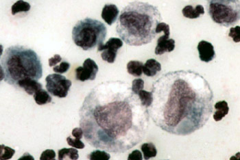
English: Color micrograph of the cytology of FIP-induced effusion. Magnification not specified; estimated to be 1000x.
Original caption: "The cytology of FIP effusion usually contains neutrophils, macrophages and lymphocytes." Image from "Feline Infectious Peritonitis: An Overview of Disease Transmission, Pathogenesis, Signs and Treatment With Emphasis on Diagnosis" ([1]) Clinical Pathology Clerkship Program |
| 날짜 | 2005년 9월 30일 (원본 올리기 일시) |
| 출처 | en.wikipedia에서 공용으로 옮겨왔습니다. |
| 저자 | The original uploader was 영어 위키백과의 Bk0. |
라이선스

|
이 파일의 저작권자는 저작권자를 명시한다는 조건에 따라 누구에게나 어떤 목적으로든지 사용할 수 있도록 허용합니다.
이 저작물의 재배포나 이차적 저작물 작성 및 상업적 이용 등이 허용됩니다. |
|
|
기존 올리기 기록
The original description page was here. All following user names refer to en.wikipedia.
- 2005-09-30 00:14 Bk0 350×234×8 (40687 bytes) Color micrograph of the cytology of [[Feline infectious peritonitis|FIP]]-induced effusion. Magnification not specified; estimated to be 1000x. Original caption: "The cytology of FIP effusion usually contains neutrophils, macrophages and lymphocytes." I
파일 역사
날짜/시간 링크를 클릭하면 해당 시간의 파일을 볼 수 있습니다.
| 날짜/시간 | 섬네일 | 크기 | 사용자 | 설명 | |
|---|---|---|---|---|---|
| 현재 | 2007년 12월 30일 (일) 03:53 | | 350 × 234 (40 KB) | Euthygenes | {{Information |Description={{en|Color micrograph of the cytology of FIP-induced effusion. Magnification not specified; estimated to be 1000x. Original caption: "The cytology of FIP effusion usually contains neutrophi |
이 파일을 사용하는 문서
다음 문서 1개가 이 파일을 사용하고 있습니다:
이 파일을 사용하고 있는 모든 위키의 문서 목록
다음 위키에서 이 파일을 사용하고 있습니다:
- el.wikipedia.org에서 이 파일을 사용하고 있는 문서 목록
- en.wikipedia.org에서 이 파일을 사용하고 있는 문서 목록
- et.wikipedia.org에서 이 파일을 사용하고 있는 문서 목록
- fr.wikipedia.org에서 이 파일을 사용하고 있는 문서 목록
- hu.wikipedia.org에서 이 파일을 사용하고 있는 문서 목록
- tr.wikipedia.org에서 이 파일을 사용하고 있는 문서 목록
- zh.wikipedia.org에서 이 파일을 사용하고 있는 문서 목록