파일:Determinants of Gastric Acid Secretion.svg

SVG 파일의 PNG 형식의 미리보기 크기: 800 × 550 픽셀. 다른 해상도: 320 × 220 픽셀 | 640 × 440 픽셀 | 1,024 × 704 픽셀 | 1,280 × 880 픽셀 | 2,560 × 1,760 픽셀 | 1,206 × 829 픽셀
원본 파일 (SVG 파일, 실제 크기 1,206 × 829 픽셀, 파일 크기: 741 KB)
파일 설명
| 설명 |
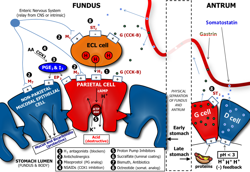
English: There are several neuronal, endocrine, and paracrine determinants of gastric acid secretion. Parietal cells in the fundus and body of the stomach are responsible for secretion of acid into the stomach. Major receptors on parietal cells that trigger acid release when activated include the M3 muscarinic, the H2 histaminic, and the CCK-B or gastrin receptor. The respective ligands for these receptors are acetylcholine, histamine, and gastrin. Acetylcholine is released from enteric neurons, histamine is released from enterochromaffin-like cells, and gastrin is released from G cells which are generally found in the antrum, or lower portion, of the stomach. Enterochromaffin-like (ECL) cells are generally found below the epithelial cell layer of the stomach lumen. ECL cells express M3 muscarinic, ST2 somatostatin, and CCK-B gastrin receptors. While M3 and CCK-B receptors trigger histamine release when activated, the ST2 somatostatin receptor inhibits histamine release. Thus, gastrin is pro-acid, and somatostatin is anti-acid secretion. Both somatostatin and gastrin are released primarily from cells in the antrum. G cells secrete gastrin, and D cells secrete somatostatin. D cells have a regulatory role over G cells in that the somatostatin that they release has an inhibitory influence on gastrin secretion from G cells. This serves to reduce futile simultaneous pro-acid and anti-acid signals to the fundus and body of the stomach. A major trigger for gastrin release is the presence of protein in the antrum: this serves to enhance acid secretion, which is necessary for the activity of pepsin, a major digestive enzyme in the stomach which is important for protein breakdown. In addition, D cells are also subject to regulation by gastric luminal contents. In particular, D cells release more somatostatin when the antral pH is low (i.e., when a highly acidic condition exists). This serves as a negative feedback when acid levels are high, as somatostatin will be secreted, enter the gastric bloodstream, and signal to ECL cells to reduce histamine release. Reduced histamine release will, in turn, reduce acid secretion from parietal cells. Finally, our gastric cells would be susceptible to damage from acid and pepsin if they had no protection. Protection of the gastric mucosal cells is in two forms, a physical barrier of viscous mucus, and a chemical defense of bicarbonate, which neutralizes acid. Protective prostaglandins, i.e., PGE2 and PGI2 activate EP3 receptors on non-parietal mucosal cells to enhance mucus and bicarbonate secretion. At the same time, EP3 receptors are activated on parietal cells to inhibit acid secretion. This is primarily a result of EP3's coupling to the G-protein, Gi, which reduces cyclic adenosine monophosphate (cAMP) levels. The proton pump in parietal cells is an exchanger of potassium and protons. Many portions of the physiology of gastric acid secretion are influenced by drugs, especially those used to treat peptic ulcer disease (PUD) and gastroesophageal reflux disease (GERD). The drug classes and sites of action are denoted by numbers. Note: non-steroidal anti-inflammatory drugs (NSAIDs) such as aspirin and ibuprofen inhibit the production of protective prostaglandins by inhibiting cyclooxygenase 1 (COX1). For this reason, NSAIDs can promote ulcer formation, and their antiplatelet effects also make patients more susceptible to excessive bleeding if a gastric ulcer becomes severe. |
| 날짜 | |
| 출처 | 자작 |
| 저자 | Adam L. VanWert, Pharm.D., Ph.D. |
| 다른 버전 | 이 파일은 다음으로 파생됨: Determinants of Gastric Acid Secretion Edit.svg |
라이선스
나는 아래 작품의 저작권자로서, 이 저작물을 다음과 같은 라이선스로 배포합니다:
이 파일은 크리에이티브 커먼즈 저작자표시 3.0 Unported 라이선스로 배포됩니다.
- 이용자는 다음의 권리를 갖습니다:
- 공유 및 이용 – 저작물의 복제, 배포, 전시, 공연 및 공중송신
- 재창작 – 저작물의 개작, 수정, 2차적저작물 창작
- 다음과 같은 조건을 따라야 합니다:
- 저작자표시 – 적절한 저작자 표시를 제공하고, 라이센스에 대한 링크를 제공하고, 변경사항이 있는지를 표시해야 합니다. 당신은 합리적인 방식으로 표시할 수 있지만, 어떤 방식으로든 사용권 허가자가 당신 또는 당신의 사용을 지지하는 방식으로 표시할 수 없습니다.
파일 역사
날짜/시간 링크를 클릭하면 해당 시간의 파일을 볼 수 있습니다.
| 날짜/시간 | 섬네일 | 크기 | 사용자 | 설명 | |
|---|---|---|---|---|---|
| 현재 | 2011년 1월 16일 (일) 20:03 | | 1,206 × 829 (741 KB) | Vanwa71 | .. |
| 2011년 1월 16일 (일) 19:57 |  | 1,206 × 829 (486 KB) | Vanwa71 | Reverted to version as of 10:39, 16 January 2011 | |
| 2011년 1월 16일 (일) 19:55 |  | 1,206 × 829 (1.33 MB) | Vanwa71 | Linked instead of embedded new graphics last time. | |
| 2011년 1월 16일 (일) 19:39 |  | 1,206 × 829 (486 KB) | Vanwa71 | Reverted to version as of 06:03, 16 January 2011 | |
| 2011년 1월 16일 (일) 19:37 |  | 1,206 × 829 (437 KB) | Vanwa71 | cool effects | |
| 2011년 1월 16일 (일) 15:03 |  | 1,206 × 829 (486 KB) | Vanwa71 | Small detail edits. | |
| 2011년 1월 16일 (일) 14:57 |  | 1,206 × 829 (484 KB) | Vanwa71 | Fixed gastrin and somatostatin flaws | |
| 2011년 1월 16일 (일) 14:14 |  | 1,206 × 829 (472 KB) | Vanwa71 | Forgot categories. | |
| 2011년 1월 16일 (일) 14:11 |  | 1,206 × 829 (472 KB) | Vanwa71 | I finally started using my head, and converted all the text to outlines to keep it exactly as I wanted it. | |
| 2011년 1월 16일 (일) 14:05 |  | 1,206 × 829 (214 KB) | Vanwa71 | Please bear with me, I finally looked up websafe fonts instead of trial and error. Let's hope this works. |
이 파일을 사용하는 문서
다음 문서 1개가 이 파일을 사용하고 있습니다:
이 파일을 사용하고 있는 모든 위키의 문서 목록
다음 위키에서 이 파일을 사용하고 있습니다:
- el.wikipedia.org에서 이 파일을 사용하고 있는 문서 목록
- en.wikipedia.org에서 이 파일을 사용하고 있는 문서 목록
- fr.wikipedia.org에서 이 파일을 사용하고 있는 문서 목록
- hi.wikipedia.org에서 이 파일을 사용하고 있는 문서 목록
- id.wikipedia.org에서 이 파일을 사용하고 있는 문서 목록
- ja.wikipedia.org에서 이 파일을 사용하고 있는 문서 목록
- sq.wikipedia.org에서 이 파일을 사용하고 있는 문서 목록
- te.wikipedia.org에서 이 파일을 사용하고 있는 문서 목록
- zh.wikipedia.org에서 이 파일을 사용하고 있는 문서 목록
- zh.wikibooks.org에서 이 파일을 사용하고 있는 문서 목록