파일:Neutrophil with anthrax copy.jpg
미리 보기 크기: 575 × 600 픽셀 다른 해상도: 230 × 240 픽셀 | 460 × 480 픽셀 | 736 × 768 픽셀 | 982 × 1,024 픽셀 | 2,304 × 2,403 픽셀
원본 파일 (2,304 × 2,403 픽셀, 파일 크기: 2.28 MB, MIME 종류: image/jpeg)
|

This image was selected as picture of the day on Vietnamese Wikipedia.
|
| This image was selected as a picture of the week on the Persian Wikipedia for the 16째 week, 2011. |
파일 설명
| 설명 |
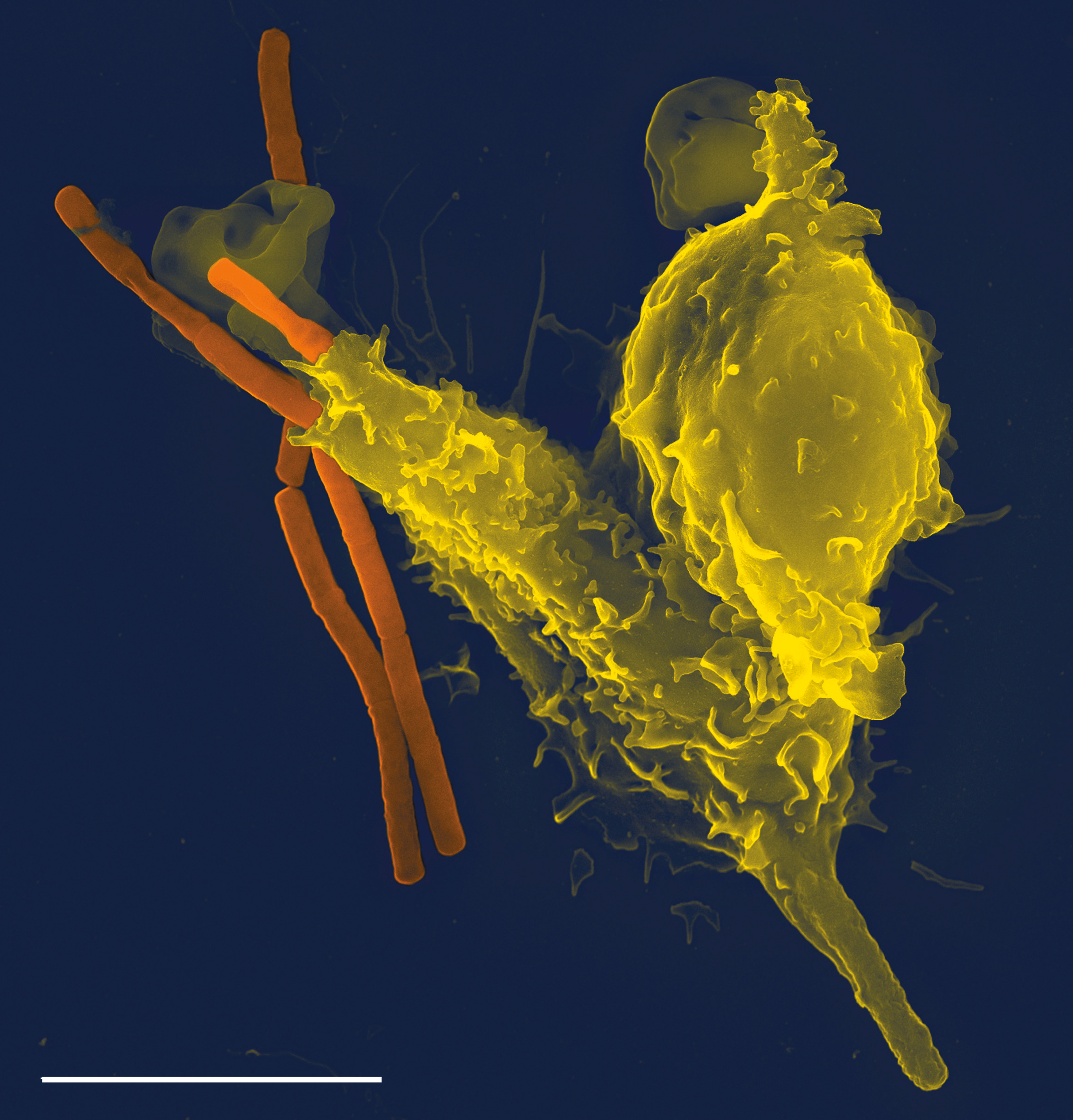
العربية: صورة بالمجهر الإلكتروني الماسح لخلية مُتعادلة (أصفر) تبتلع بكتيريا الجَمْرَة الخبيثة (برتقالي)
Deutsch: Eine REM-Aufnahme eines neutrophilen Granulozyten (gelb) beim Aufnehmen von Anthrax-Bakterien (orange).
Maßstab: Der weiße Strich links unten ist 5 Mikrometer lang. English: Neutrophil engulfing anthrax bacteria, taken with a Leo 1550 scanning electron microscope. Scale bar is 5 micrometers.
Español: Neutrófilo (en amarillo) engullendo una bacteria de carbunco o ántrax (en naranja). Fotografía tomada con un microscopio de electrones Leo 1550. La escala son 5 micrómetros.
Français : Un neutrophile (en jaune) phagocytant des bacilles du charbon (en orange). Photo prise avec un microscope electronique. Echelle: 5 microns.
Nederlands: Elektronenmicroscopieopname van fagocytose tussen een fagocyt (geel) en miltvuurbacteriën (oranje)
Tiếng Việt: Hình ảnh chụp bằng kính hiển vi điện tử quét cho thấy vi khuẩn Salmonella (nhuộm màu đỏ) xâm nhập vào tế bào của con người. Salmonella là tác nhân gây ra sốt thương hàn và ngộ độc thực phẩm.
中文(臺灣):嗜中性白血球正在吞噬鼠疫桿菌。此電子顯微鏡影像由Leo 1550掃描式電顯拍攝。左下角比例尺為5微米。 |
| 날짜 | |
| 출처 | (November 2005). "Neutrophil engulfing Bacillus anthracis". PLoS Pathogens 1 (3): Cover page. DOI:10.1371. Archived from the original on 2015-01-09. Retrieved on 2009-01-04. |
| 저자 | Volker Brinkmann |
| 저작권 (이 파일을 인용하기) |
All PLoS content released under CC-BY license.[1] 이 파일은 크리에이티브 커먼즈 저작자표시 2.5 일반 라이선스로 배포됩니다.
|
기존 올리기 기록
(All user names refer to en.wikipedia)
- 2007-03-23 04:24 TimVickers 2304×2403×8 (2243510 bytes) Neutrophil engulfing anthrax bacteria. Cover credit: The micrograph was taken by Volker Brinkmann with a Leo 1550 scanning electron microscope. Taken from PLoS Pathogens Vol. 1(3) November 2005 --
파일 역사
날짜/시간 링크를 클릭하면 해당 시간의 파일을 볼 수 있습니다.
| 날짜/시간 | 섬네일 | 크기 | 사용자 | 설명 | |
|---|---|---|---|---|---|
| 현재 | 2007년 9월 26일 (수) 01:00 | | 2,304 × 2,403 (2.28 MB) | TimVickers | Neutrophils are the most abundant white blood cell and the first line of defense against invading microbes. This scanning electron micrograph shows a neutrophil (yellow) engulfing rods of Bacillus anthracis (orange), the etiological agent of anthrax (see |
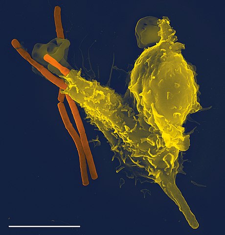
| 2007년 5월 15일 (화) 00:38 |  | 2,304 × 2,403 (2.14 MB) | Kauczuk | {{Information |Description=Neutrophil engulfing anthrax bacteria. Cover credit: The micrograph was taken by Volker Brinkmann with a Leo 1550 scanning electron microscope. Taken from PLoS Pathogens Vol. 1(3) November 2005 |Source=Originally from [http://en |
이 파일을 사용하는 문서
다음 문서 7개가 이 파일을 사용하고 있습니다:
이 파일을 사용하고 있는 모든 위키의 문서 목록
다음 위키에서 이 파일을 사용하고 있습니다:
- af.wikipedia.org에서 이 파일을 사용하고 있는 문서 목록
- als.wikipedia.org에서 이 파일을 사용하고 있는 문서 목록
- an.wikipedia.org에서 이 파일을 사용하고 있는 문서 목록
- ar.wikipedia.org에서 이 파일을 사용하고 있는 문서 목록
- جهاز مناعي
- بوابة:طب/صورة مختارة
- خلية متعادلة
- خلية بلعمية
- بوابة:علم الأحياء/صورة مختارة/أرشيف
- ويكيبيديا:صور مختارة/علوم/علم الأحياء
- بوابة:طب/صورة مختارة/4
- ويكيبيديا:ترشيحات الصور المختارة/خلية متعادلة
- ويكيبيديا:صورة اليوم المختارة/نوفمبر 2020
- قالب:صورة اليوم المختارة/2020-11-29
- بوابة:علم الأحياء/صورة مختارة/31
- ويكيبيديا:صورة اليوم المختارة/أكتوبر 2021
- قالب:صورة اليوم المختارة/2021-10-24
- arz.wikipedia.org에서 이 파일을 사용하고 있는 문서 목록
- ast.wikipedia.org에서 이 파일을 사용하고 있는 문서 목록
- as.wikipedia.org에서 이 파일을 사용하고 있는 문서 목록
- az.wikipedia.org에서 이 파일을 사용하고 있는 문서 목록
- bat-smg.wikipedia.org에서 이 파일을 사용하고 있는 문서 목록
- ba.wikipedia.org에서 이 파일을 사용하고 있는 문서 목록
- be-tarask.wikipedia.org에서 이 파일을 사용하고 있는 문서 목록
- bg.wikipedia.org에서 이 파일을 사용하고 있는 문서 목록
- bn.wikipedia.org에서 이 파일을 사용하고 있는 문서 목록
- bs.wikipedia.org에서 이 파일을 사용하고 있는 문서 목록
- ca.wikipedia.org에서 이 파일을 사용하고 있는 문서 목록
- Viquipèdia:Articles seleccionats/maig
- Plantilla:Article maig 20
- Viquipèdia:Articles seleccionats/setembre
- Plantilla:Article setembre 1
- Neutròfil
- Sistema immunitari
- Fagòcit
- Portal:Immunologia/Imatges
- Plantilla:Imatge immunològica del mes 0
- Icosoma
- Viquipèdia:Imatge del dia/2015
- Portada/article maig 20
- Portada/article setembre 1
- ckb.wikipedia.org에서 이 파일을 사용하고 있는 문서 목록
- crh.wikipedia.org에서 이 파일을 사용하고 있는 문서 목록
- cs.wikipedia.org에서 이 파일을 사용하고 있는 문서 목록
이 파일의 더 많은 사용 내역을 봅니다.