오른관상동맥
심장의 관상순환에서 오른관상동맥(right coronary artery, RCA) 또는 오른심장동맥, 우관상동맥(右冠狀動脈)은 대동맥판의 오른쪽 첨판에 존재하는 오른쪽 대동맥굴에서 시작되는 동맥이다.[1][2] 이후에는 오른쪽 관상고랑을 따라 이동한다.[1][3] 심장의 오른쪽과 심실사이막에 혈액을 공급한다.[2][4]
| 오른관상동맥 | |
|---|---|
 심장의 관상순환. 오른관상동맥이 표시됨. | |
| 정보 | |
| 위치 | 심장 |
| 혈액 공급 | 우심방, 우심실 좌심실의 25-35% |
| 식별자 | |
| 라틴어 | arteria coronaria dextra |
| 영어 | right coronary artery |
| TA98 | A12.2.03.101 |
| TA2 | 4131 |
| FMA | 50039 |
구조 편집
오른관상동맥은 대동맥판 위의 오른쪽 대동맥굴 위에서 시작된다.[1][2] 이후 오른쪽 관상고랑(오른쪽 방실사이고랑)을 따라 관상고랑과 뒤심실사이고랑이 만나는 지점까지 주행한다.[1][3] 뒤심실사이동맥, 오른모서리가지, 굴심방결절동맥 등의 많은 가지를 낸다.[5]
분절 편집
- 몸쪽: 오른관상동맥이 시작하는 지점부터 심장 오른쪽 경계까지 가는 길이의 절반 정도를 차지한다.[6][7]
- 중간: 몸쪽 부분이 끝나는 지점에서 오른쪽 경계까지의 부분.[6][7]
- 먼쪽: 중간 부분부터 뒤심실사이동맥 시작 지점까지의 부분. 뒤심실사이고랑은 심장 바닥에서 관상고랑과 만나며 이 지점에서 뒤심실사이동맥이 시작된다.[6][7]
변이 편집
환자의 약 80%(오른쪽 우세, right dominant인 경우)에서 오른관상동맥은 뒤심실사이동맥(뒤내림동맥)을 낸다. 다른 20%의 경우(왼쪽 우세, left dominant거나 공동 우세, codominant인 경우)에서 뒤심실사이동맥은 왼관상동맥 휘돌이가지에서 의해 나오거나 오른관상동맥과 휘돌이가지 양쪽 모두에 의해 형성된다.[8] 오른관상동맥은 심장의 아래쪽 벽, 심실사이막, 뒤안쪽 꼭지근에 혈액을 공급한다.
또한 오른관상동맥은 60%의 경우에서 굴심방결절동맥을 낸다. 나머지 40%에서는 휘돌이가지가 굴심방결절동맥을 낸다.
드물기는 하지만 왼쪽 대동맥굴에서 나오는 경우처럼 오른관상동맥의 몇 가지 비정상적인 경로가 기술된 바 있다.[9]
기능 편집
오른관상동맥은 우심방, 우심실, 심실사이막의 뒤쪽 3분의 1과 아래쪽 끝에 산소가 풍부한 혈액을 공급한다.[2][4] 좌심실의 25~35%에 혈액을 공급할 수도 있다.[10]
관상동맥의 공급은 크게 겹친다.[2] 오른관상동맥은 50%의 경우 왼관상동맥보다 우세하고 20%의 경우와는 대등하며 30%의 경우에서는 왼관상동맥이 더 우세하다.[2]
추가 이미지 편집
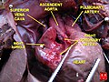
-
 오른관상동맥.
 -
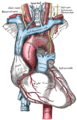 대동맥활과 그 가지.
 -
 심근 경색의 다이어그램.
 -
-
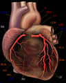 관상동맥이 표시된 인간의 심장.
 -
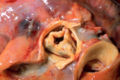
 태아 심장의 오른관상동맥.


참고 문헌 편집
- ↑ 가 나 다 라 Aggeli, Constantina; Mavrogeni, Sofia; Tousoulis, Dimitris (2018년 1월 1일), Tousoulis, Dimitris, 편집., “Chapter 3.5.1 - Non-invasive Imaging Techniques in Coronary Artery Disease”, 《Coronary Artery Disease》 (영어) (Academic Press), 337–358쪽, doi:10.1016/b978-0-12-811908-2.00017-9, ISBN 978-0-12-811908-2, 2020년 11월 20일에 확인함
- ↑ 가 나 다 라 마 바 Pappano, Achilles J.; Gil Wier, Withrow (2013년 1월 1일), Pappano, Achilles J.; Gil Wier, Withrow, 편집., “11 - Coronary Circulation”, 《Cardiovascular Physiology (Tenth Edition)》 (영어) (Philadelphia: Elsevier), 223–236쪽, doi:10.1016/b978-0-323-08697-4.00011-3, ISBN 978-0-323-08697-4, 2020년 11월 20일에 확인함
- ↑ 가 나 Sivananthan, M. (2018년 1월 1일), 〈Coronary Anatomy〉, Vasan, Ramachandran S.; Sawyer, Douglas B., 《Encyclopedia of Cardiovascular Research and Medicine》 (영어), Oxford: Elsevier, 691–699쪽, doi:10.1016/b978-0-12-809657-4.99738-2, ISBN 978-0-12-805154-2, 2020년 11월 20일에 확인함
- ↑ 가 나 Schipper, Paul; Sukumar, Mithran; Mayberry, John C. (2008년 1월 1일), Asensio, JUAN A.; Trunkey, DONALD D., 편집., “Pertinent Surgical Anatomy of the Thorax and Mediastinum”, 《Current Therapy of Trauma and Surgical Critical Care》 (영어) (Philadelphia: Mosby), 227–251쪽, doi:10.1016/b978-0-323-04418-9.50037-0, ISBN 978-0-323-04418-9, 2020년 11월 20일에 확인함
- ↑ Antonopoulos, Alexios S.; Siasos, Gerasimos; Antoniades, Charalambos; Tousoulis, Dimitris (2018년 1월 1일), Tousoulis, Dimitris, 편집., “Chapter 2.1 - Functional Anatomy”, 《Coronary Artery Disease》 (영어) (Academic Press), 121–126쪽, doi:10.1016/b978-0-12-811908-2.00008-8, ISBN 978-0-12-811908-2, 2020년 11월 20일에 확인함
- ↑ 가 나 다 Villa, AD; Sammut, E; Nair, A; Rajani, R; Bonamini, R; Chiribiri, A (2016년 6월 28일). “Coronary artery anomalies overview: The normal and the abnormal.”. 《World Journal of Radiology》 8 (6): 537–55. doi:10.4329/wjr.v8.i6.537. PMC 4919754. PMID 27358682.
- ↑ 가 나 다 Kini, S; Bis, KG; Weaver, L (June 2007). “Normal and variant coronary arterial and venous anatomy on high-resolution CT angiography.”. 《AJR. American Journal of Roentgenology》 188 (6): 1665–74. doi:10.2214/AJR.06.1295. PMID 17515392.
- ↑ Shahoud, James S.; Ambalavanan, Manoj; Tivakaran, Vijai S. (2020). 《Cardiac Dominance》. StatPearls Publishing. PMID 30725892.
- ↑ Angelini, P. (2014년 7월 15일). “Novel Imaging of Coronary Artery Anomalies to Assess Their Prevalence, the Causes of Clinical Symptoms, and the Risk of Sudden Cardiac Death”. 《Circulation: Cardiovascular Imaging》 7 (4): 747–754. doi:10.1161/CIRCIMAGING.113.000278. PMID 25027456.
- ↑ Wheeler, Derek S.; Wong, Hector R.; Shanley, Thomas P. (2014년 5월 22일). 《Pediatric Critical Care Medicine: Volume 2: Respiratory, Cardiovascular and Central Nervous Systems》 (영어). Springer. 306쪽. ISBN 978-1-4471-6356-5.