파일:Structural organization of the heart of the mosquito Anopheles gambiae - image.ppat.v08.i11.g001.png
미리 보기 크기: 600 × 600 픽셀 다른 해상도: 240 × 240 픽셀 | 480 × 480 픽셀 | 656 × 656 픽셀
원본 파일 (656 × 656 픽셀, 파일 크기: 670 KB, MIME 종류: image/png)
파일 설명
| 설명 |
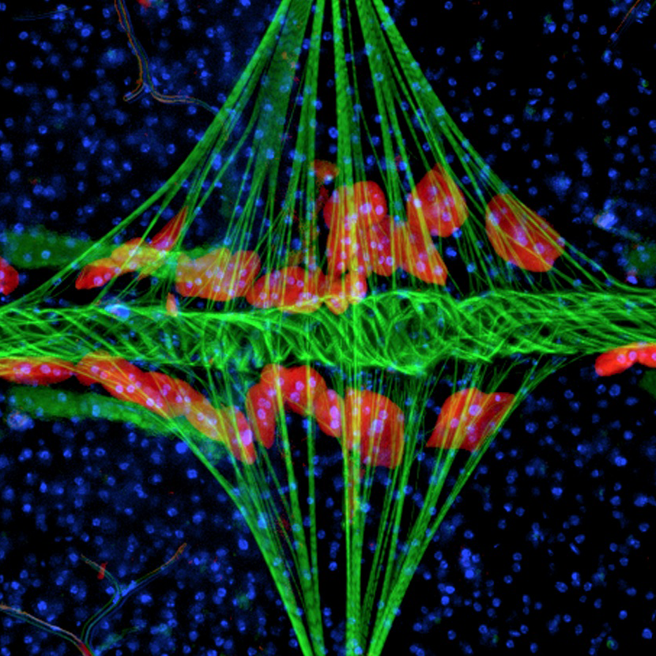
English: This fluorescence image details the structural organization of the heart of the mosquito Anopheles gambiae. The point of view is top down, with the mosquito's body lying horizontally with its head to the left (outside of the image). Muscle is labeled with phalloidin (green), and shows the tube-like heart extending horizontally across the body and the diamond shaped alary muscles projecting vertically onto the heart. The pericardial cells, labeled with 568 nm-Immunoglobulin G (red), are pinocytic cells that flank the heart. Cell nuclei are labeled with Hoechst 33342 (blue).
Image Credit: Jonas G. King and Julián F. Hillyer, Department of Biological Sciences, Vanderbilt University. Issue image, PLOS Pathogens, November 2012. |
| 날짜 | |
| 출처 | King JG, Hillyer JF (2012) Infection-Induced Interaction between the Mosquito Circulatory and Immune Systems. PLoS Pathog 8(11): e1003058. doi:10.1371/journal.ppat.1003058 |
| 저자 | King JG, Hillyer JF |
라이선스
이 파일은 크리에이티브 커먼즈 저작자표시 2.5 일반 라이선스로 배포됩니다.
- 이용자는 다음의 권리를 갖습니다:
- 공유 및 이용 – 저작물의 복제, 배포, 전시, 공연 및 공중송신
- 재창작 – 저작물의 개작, 수정, 2차적저작물 창작
- 다음과 같은 조건을 따라야 합니다:
- 저작자표시 – 적절한 저작자 표시를 제공하고, 라이센스에 대한 링크를 제공하고, 변경사항이 있는지를 표시해야 합니다. 당신은 합리적인 방식으로 표시할 수 있지만, 어떤 방식으로든 사용권 허가자가 당신 또는 당신의 사용을 지지하는 방식으로 표시할 수 없습니다.
파일 역사
날짜/시간 링크를 클릭하면 해당 시간의 파일을 볼 수 있습니다.
| 날짜/시간 | 섬네일 | 크기 | 사용자 | 설명 | |
|---|---|---|---|---|---|
| 현재 | 2015년 7월 25일 (토) 05:13 | | 656 × 656 (670 KB) | Cmdrjameson | Compressed with pngout. Reduced by 260kB (27% decrease). |
| 2012년 12월 2일 (일) 11:07 |  | 656 × 656 (930 KB) | Daniel Mietchen | User created page with UploadWizard |
이 파일을 사용하는 문서
다음 문서 2개가 이 파일을 사용하고 있습니다:
이 파일을 사용하고 있는 모든 위키의 문서 목록
다음 위키에서 이 파일을 사용하고 있습니다:
- ar.wikipedia.org에서 이 파일을 사용하고 있는 문서 목록
- bn.wikipedia.org에서 이 파일을 사용하고 있는 문서 목록
- en.wikipedia.org에서 이 파일을 사용하고 있는 문서 목록
- hy.wikipedia.org에서 이 파일을 사용하고 있는 문서 목록
- outreach.wikimedia.org에서 이 파일을 사용하고 있는 문서 목록
- si.wikipedia.org에서 이 파일을 사용하고 있는 문서 목록